![]()
What Is Diagnostic Imaging in Veterinary Medicine?
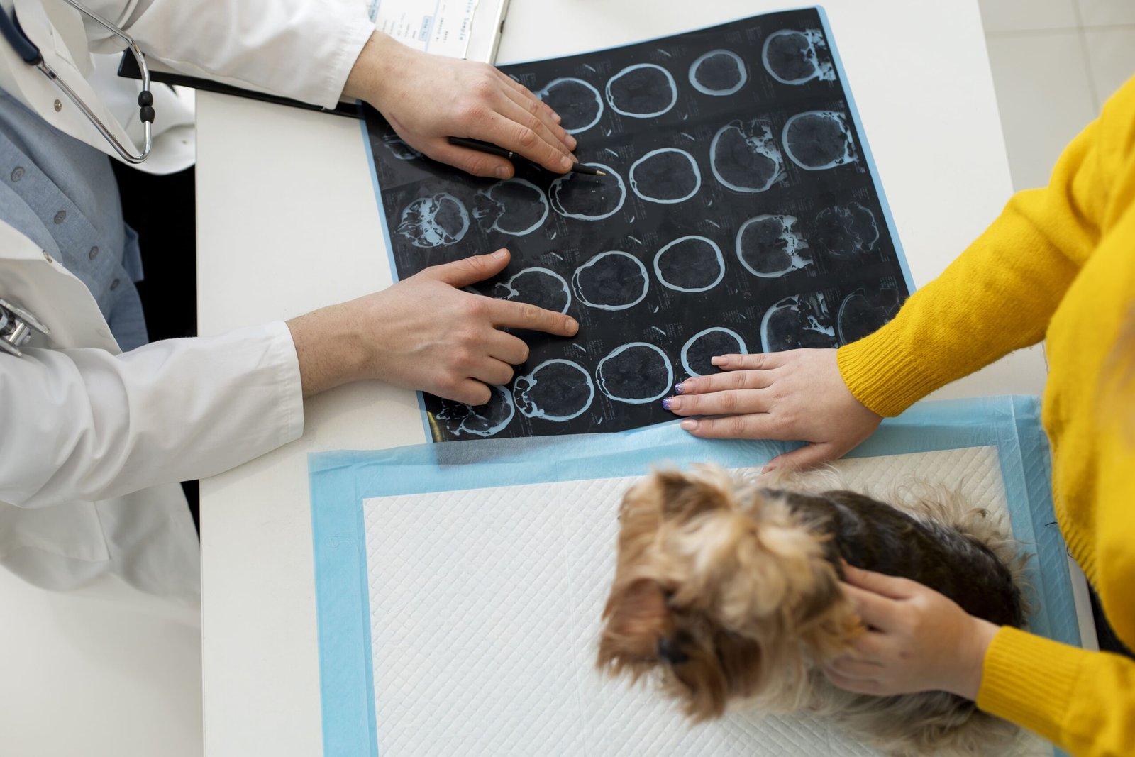
Diagnostic imaging is one of the most important tools in modern veterinary medicine. It allows veterinarians to look inside your pet’s body without the need for surgery, helping diagnose a wide range of conditions — from broken bones to organ problems. Two of the most commonly used imaging methods in pets are X-rays and ultrasounds. While both serve similar purposes, they are used in very different ways depending on the situation.
Understanding how each technique works and when it’s used can help you make better decisions about your pet’s health care.
X-rays for Pets:
X-rays (also known as radiographs) use a small dose of electromagnetic radiation to create images of the inside of your pet’s body. They’re especially useful for viewing bones, joints, and dense tissues like the lungs or heart.
Veterinarians often use X-rays to:
- Diagnose bone fractures or joint problems
- Locate foreign objects swallowed or stuck inside the body
- Detect tumors or abnormal growths
- Assess the condition of the lungs and chest cavity
X-rays are quick to perform and typically don’t require sedation unless your pet is extremely anxious or in pain. They’re widely available and usually cost less than more advanced imaging methods.
However, X-rays do have limitations. They’re not very effective at showing soft tissues like organs or muscles in detail, and they expose your pet to a small amount of radiation (though it’s generally considered safe for occasional use).
Ultrasounds for Pets:
Ultrasounds work differently. Instead of using radiation, they rely on high-frequency sound waves to create real-time images of the internal organs. This makes ultrasounds a safer option, especially for pets that need frequent imaging or are pregnant.
Veterinarians commonly use ultrasounds to:
- Examine organs like the liver, kidneys, bladder, and spleen
- Detect fluid buildup or abnormal masses
- Confirm and monitor pregnancies
- Evaluate heart function (through a special type of ultrasound called echocardiography)
Ultrasounds provide detailed visuals of soft tissue and organ structure, making them ideal for identifying internal problems that might not appear clearly on an X-ray. The procedure is painless and non-invasive, although some pets may need mild sedation to stay still during the scan.
On the downside, ultrasounds can’t effectively image bones or air-filled areas like the lungs, and they may take longer than X-rays to perform. The cost is also typically higher, depending on the complexity of the scan.
Comparing X-rays and Ultrasounds in Veterinary Diagnosis:
While both X-rays and ultrasounds are essential diagnostic tools, they serve different purposes and are chosen based on your pet’s specific condition.
X-rays are the go-to method for assessing bones, joints, and dense structures such as the chest cavity and lungs. They are fast, accessible, and affordable, making them ideal for emergencies and structural issues.
Ultrasounds, on the other hand, are the preferred choice when your veterinarian needs to look closely at organs or soft tissue structures. They offer much more detail for things like kidney disease, liver problems, or heart abnormalities and are completely free from radiation.
In some cases, veterinarians may recommend using both X-rays and ultrasounds together. For example, if a cat is vomiting and an X-ray shows something unusual in the abdomen, an ultrasound might be used to get a clearer look at the affected organs.
Ultimately, the choice between the two methods depends on what the vet is trying to investigate, your pet’s symptoms, and the level of detail required for a proper diagnosis.
What Pet Owners Should Expect:
If your veterinarian recommends imaging for your pet, you might be wondering what to expect.
Before the procedure, some ultrasounds may require your pet to fast, especially for abdominal scans. X-rays typically don’t need any preparation unless sedation is involved.
During the procedure, X-rays take only a few minutes. Your pet will be gently positioned while the machine captures images. Ultrasounds may take a bit longer and involve applying a special gel to the area being scanned. Most pets tolerate both procedures well.
Cost-wise, X-rays are generally more affordable than ultrasounds, but prices vary depending on location, clinic, and your pet’s condition. Both procedures are considered safe, with ultrasounds being completely radiation-free and X-rays involving only minimal exposure.
Conclusion: Choosing the Right Imaging for Your Pet:
Diagnostic imaging is a powerful way for veterinarians to diagnose health issues in pets without invasive procedures. X-rays are excellent for viewing bones and dense tissues, while ultrasounds offer detailed views of soft organs and internal systems. Both have their strengths, and the right choice depends on your pet’s symptoms and the vet’s clinical judgment.
As a pet owner, knowing the differences between these tools can help you feel more confident and prepared during your pet’s health journey. Always follow your veterinarian’s advice—they will choose the imaging method that provides the clearest and safest results for your furry companion.